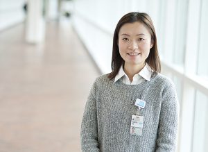
Congratulations to biomedical engineering graduate student Xiaoying Cai for being selected as one of seven fellows by the American Heart Association and the Children’s Heart Foundation for a joint funding venture in congenital heart defect research. Cai works in Dr. Fred Epstein’s lab and was the only predoctoral fellow selected; the other six awardees have already earned their MDs or PhDs.
Cai is working on a new method to use magnetic resonance imaging to help children born with single ventricle defects, with a goal of locating regions of the heart best suited to interventions like advanced pacemakers. Her clinical collaborator is Dr. Mark Fogel, a colleague at the Children’s Hospital of Pennsylvania.
Please join us in congratulating Xiaoying on this award!
Here is a more detailed explanation of her research, from her application:
What is the major problem being addressed by this study?
This proposal addresses the problem of how to assess and potentially improve treatment for patients who are born with single ventricle defects and who develop cardiac dysfunction, even after the reparative Fontan reconstruction operation. First, we propose to develop a free-breathing imaging method so that we can better image cardiac function in young subjects to quantify dyssynchronous contraction (dyssynchrony) and detect late-activating regions of the heart. Second, we propose to assess dyssynchrony and myocardial scar using the new imaging method along with the standard scar imaging protocol to investigate whether MRI can detect regions of the heart that are both late-activated and do not have scar. This study might demonstrate MRI as a powerful tool to optimize management and treatment.
What specific questions are you asking and how will you attempt to answer them?
We would like to know the severity and prevalence of cardiac dyssynchrony in single ventricle patients. First, we will develop a repeatable free-breathing strain imaging method using advanced MRI engineering. Next we will apply this technique to image single ventricle patients and quantify dyssynchrony. By using imaging both strain and scar using MRI, we will determine whether single ventricle patients have locations in their hearts that are both late activating and free of scar, as such regions would be optimal target locations for implementing advanced pacemaker devices to resynchronize the heart and improve cardiac function.
What is the long-term biomedical significance of your work, particularly as it pertains to the cardiovascular area? What major therapeutic advance(s) do you anticipate that it will lead to?
The successful completion of this work will provide improved imaging methods to quantify cardiac dyssynchrony in young subjects and potentially lead to better therapies for patients who have undergone surgical repair of congenital heart disease.
Filed Under: Education, Honors & Awards, Research


Comments